Science
University of Oxford develops technology to reconstruct label-free molecular maps Results match fluorescence staining, with development also trackable

A technology has been developed to reconstruct 3D molecular maps of neural stem cell clusters (neural organoids) without staining or sectioning.
A research team led by Molly Stevens, a professor at the University of Oxford in the United Kingdom, said on the 1st that it had developed a platform that combines deep learning and Raman spectroscopy to analyze the molecular composition of neural organoids without labels. The findings were published in the international journal Science Advances.
Raman spectroscopy uses a laser directed at a sample to read scattering signals that differ among molecules. However, spectra measured at a single point contain signals from multiple molecules, making them difficult to interpret in complex biological samples.
The team used a custom-developed autoencoder neural network to separate molecule-specific signals from mixed spectra and calculate the abundance of each component at each point.
To verify the method’s accuracy, the team compared images reconstructed with deep learning pixel by pixel with results from conventional immunofluorescence staining. The cell nuclei and cell membranes showed high concordance with DAPI and DiO fluorescence signals, respectively. The apical membrane region of neural rosettes, a characteristic structure of neural organoids, was also reproduced in nearly the same location and shape as in fluorescence images.
When the team increased the resolution to 1 micrometer (㎛) and imaged entire organoids without damaging them, it could distinguish cell nuclei, cell membrane boundaries and intracellular lipid droplets. Layer-by-layer scanning and 3D reconstruction also revealed the three-dimensional structure of neural rosettes in its entirety for the first time.
The team also tracked development by observing organoids at five time points from day 5 to day 40 of culture. From days 5 to 10, cell proliferation was active and nucleic acid signals peaked. After day 20, lipid signals increased markedly in step with the periods of neurite growth and synapse formation. The method recorded molecular-level developmental changes without using antibodies.
The team said the technology could be used to track drug distribution, screen phenotypes in disease models and manage stem cell quality. Because Raman spectroscopy is compatible with live cell cultures, the team expects real-time molecular observation to be possible in the future.
Biobook Team
biobookmedia@biobook.co.kr
Roche and Zealand’s Amylin Drug Shows Limited Glucose-Lowering Effect, Potentially Narrowing Its Role to Maintenance Therapy

HLB Makes Fourth Attempt to Win Liver Cancer Drug Approval; Manufacturing Inspection Risks Remain

HLB Submits FDA Resubmission for Liver Cancer First-Line Treatment

Nature Cell Wins First-Instance Ruling on Jointstem … “Order for Reconsideration, Not Approval”
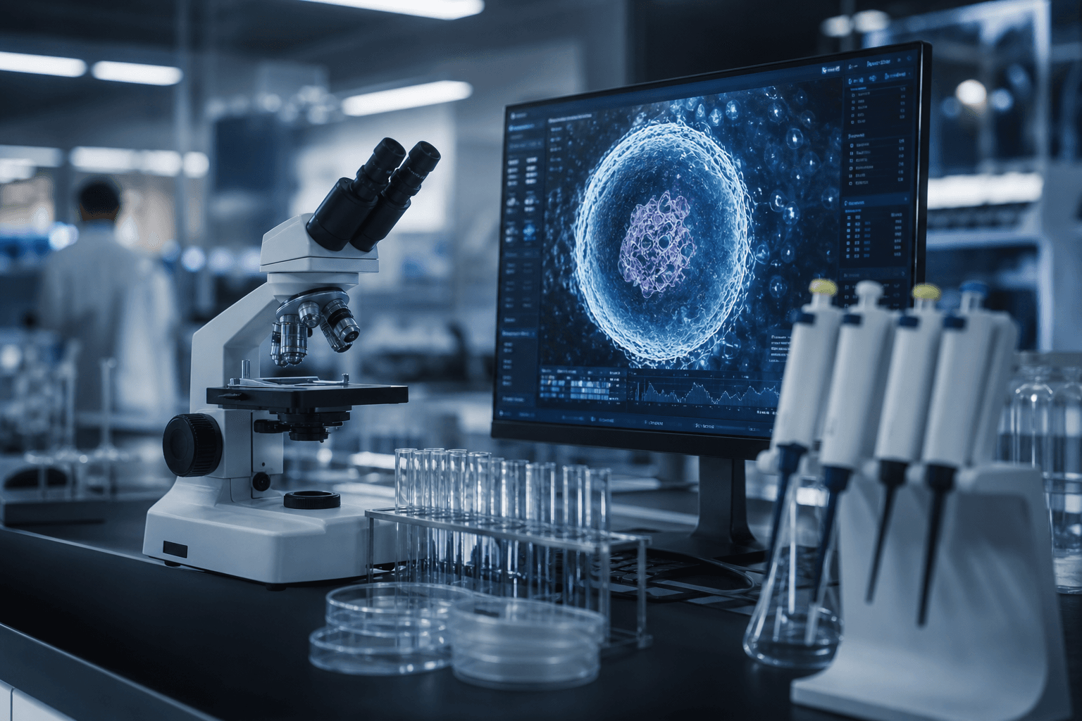
Biohub’s $2.4 Billion ‘Virtual Cell’ Project Gives Big Tech 1-Year Exclusive Data Access

Korean Biotechs Target DMOADs for Cartilage Regeneration in Race for Global First
Merck’s $1.3 trillion eye disease drug raises safety and efficacy concerns: “Limited competitiveness”
CHA Biotech Holds 24.23% Stake in CMG Pharmaceutical as Share Consolidation Reduces Its Share Count
Rural Development Administration Develops Two Molecular Markers to Identify Leaf Splitting in Squash